On the 10th of July this year, a 10 year old brood mare was referred to us by another practice for swelling and heat in the calcaneal bursa area of the right hind (at the back of the hock). She was very lame (4/5) and painful on palpation of that area. There had been history of a tiny wound on the inside of the hock but when she came in, there was no evidence of open wounds.
When she arrived, the mare was sedated for a thorough ultrasound scan of the hock area, which showed evidence of severe inflammation of the sub-tendineous calcaneal bursa. This bursa is located between the superficial flexor tendon, the gastrocnemius muscle and the calcaneus, it prevents the tendon from rubbing against the bone surface.
This is a pathology that should be taken very seriously, especially in the chronic stage. The prognosis is guarded and aggressive treatment is required in order to acquire healing. We decided to perform bursoscopy in order to flush the calcaneal bursa so that the inflammation could settle and the pressure would be taken off the surrounding tissues. We also performed IVRA (intravenous regional antibiosis) in the affected limb during surgery. In order to do this, a tourniquet was placed above the hock and antibiotic was injected into the metatarsal vein. By leaving the tourniquet in place for 10-15 minutes, the antibiotic can diffuse locally and obtain high concentrations at the infection site.
The surgery went very well and the mare is sound after about a month of recovery despite the poor prognosis.

On this ultrasound image distension of the bursa is visible, containing strands of fibrin

On this image you can see the difference between the affected bursa (right hind) and the normal limb (left hind). On the image of the right hind the superficial flexor tendon is visible at the top of the image and the gastrocnemius tendon is visible at the bottom, separated by the droplet shaped inflamed bursa.
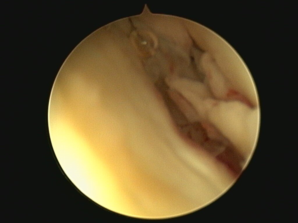
Bursoscopy image from during the surgery looking inside the calcaneal bursa. The red fibrous aspect of the inside of the bursa is an indication of severe inflammation.

Bursoscopy image where you can see the fibrous/flaky content of the inflamed bursa. This was debrided and flushed out.
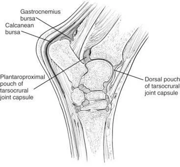
Anatomy of the hock of the horse. “calcanean bursa” indicates the subtendineous bursa we performed surgery on (situated between the superficial flexor tendon and the gastrocnemius muscle.